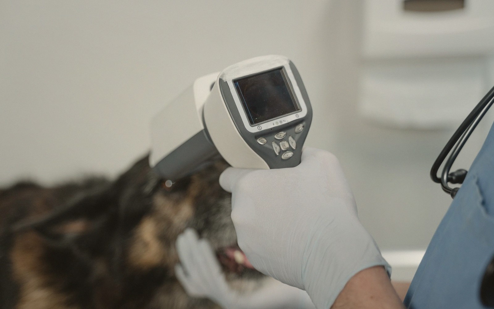
X-Ray and Ultrasound, Compared
Two imaging tools, two different questions — and the reasons your vet might reach for both.
What Each One Is Actually Showing
An X-ray is a map of density. The beam passes through the body and lands on a detector; dense structures such as bone absorb most of the radiation and appear white, while air appears almost black, and soft tissues fall somewhere in between. That contrast is exactly what makes X-ray the right tool for fractures, foreign objects lodged in the gut, and the size and shape of organs — a swollen heart silhouette, fluid in the chest cavity, a bone fragment that should not be there. The image is produced in seconds, reads at a glance, and requires nothing more exotic than the patient lying still.
Ultrasound works entirely differently. A probe sends high-frequency sound waves into the body and listens for the echoes that bounce back; the machine converts those echoes into a live image on screen. Because it captures movement in real time — a heartbeat, a bladder filling and emptying, blood flowing through a vessel — ultrasound answers questions that a still X-ray simply cannot. It can distinguish a fluid-filled cyst from a solid mass. It shows the internal architecture of the liver, spleen and kidneys in detail. And for cardiology work — assessing how well the heart muscle is contracting, measuring wall thickness, watching valve function — it is irreplaceable.
The simplest way to hold both in mind: X-ray shows structure and position; ultrasound shows texture, movement and what is inside.
No doses and no home remedies here: anything you give an animal should come from your own vet, for that animal.
Why Ultrasound Often Needs Sedation — and X-Ray Mostly Does Not
A standard X-ray takes a fraction of a second. The main challenge is keeping the animal still long enough for the image to come out sharp rather than blurred — which is usually achieved with gentle positioning, sometimes a mild sedative, and occasionally a nurse holding the patient in place. For a calm dog or cat, it is often no more stressful than being held on an examination table. Rabbits and small mammals require more care, because the stress of restraint can itself be dangerous in some species, but the procedure itself is brief.
Ultrasound is more demanding, for one practical reason: the probe must maintain close, continuous contact with the skin. Fur traps air, and air blocks sound waves completely, so the relevant area has to be clipped — sometimes a surprisingly large patch — and acoustic gel applied. The animal then has to stay genuinely still, often on its back or side, while the sonographer moves the probe slowly across the abdomen or chest. A cooperative patient can manage this awake. An anxious one, or any patient where the vet needs a detailed look at a small or deep structure, will very often need sedation — not a full anaesthetic, but enough chemical relaxation to prevent movement and keep the image readable. Sedation also makes the experience less stressful for the animal, which matters.
This is worth knowing in advance because sedation adds to the visit: there is a pre-procedure check, a waiting period while it takes effect, and a recovery period before the animal goes home. A trip that was booked as a quick scan can become a half-day appointment.
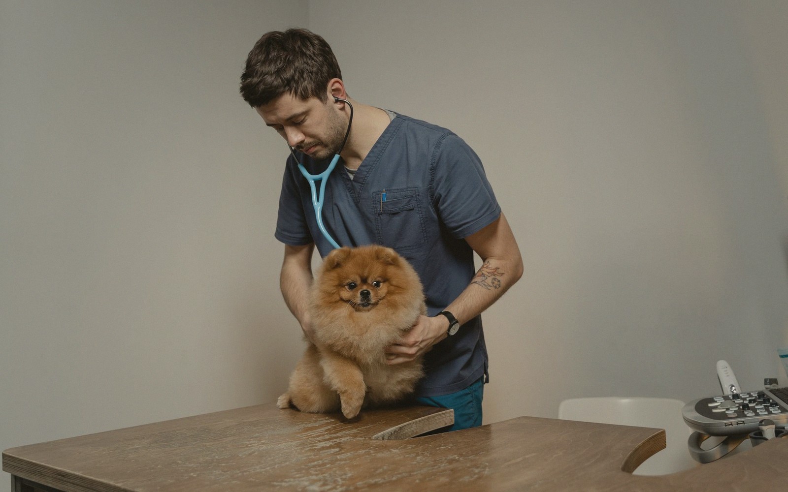
When a Vet Uses Both
The two modalities complement rather than compete. A dog that swallowed something it should not have might have an X-ray first — to confirm there is an object, and to see where it sits in the gut. If the X-ray is inconclusive (soft objects such as fabric show little contrast), an ultrasound follows to look in more detail. A cat with weight loss and a vague abdominal mass might have X-ray chest views taken first to check for obvious spread to the lungs, then an abdominal ultrasound to characterise the mass itself. In cardiac cases, chest X-rays and echocardiography (cardiac ultrasound) are routinely done together because each gives information the other cannot.
Specialist centres carry more advanced equipment — CT scanners and MRI machines — that build three-dimensional images from hundreds of slices and are used for complex neurological, orthopaedic or oncological cases. These exist further along the referral pathway; most first-opinion practices rely on X-ray and ultrasound as their everyday imaging pair, and between them those two tools answer the majority of questions that arise.
If your vet proposes imaging, it is entirely reasonable to ask which modality and why, whether sedation is likely, and how quickly results will be available. Some practices can read images on the day; others send them to a specialist radiologist for a written report, which takes longer. Knowing this before you leave with your pet means no anxious wait wondering if no news is good news.