What a Dental Actually Involves
Scale, polish, probe and often x-ray — a dental is a proper procedure, and understanding what happens under anaesthetic makes the whole thing less daunting.
What the vet is actually doing
A dental in veterinary medicine is not a hygiene visit. It is a clinical procedure carried out under general anaesthetic, and it covers a lot of ground in a single session — cleaning that cannot safely be done on a conscious animal, assessment of every tooth, and any extractions or treatments the examination reveals are necessary.
The anaesthetic is not optional or added for convenience. Scaling plaque and tartar from tooth surfaces — particularly below the gum line — requires precision instruments and complete stillness. An awake animal cannot provide that stillness safely, and the gum pockets where disease hides are inaccessible without it. Anaesthetic-free dental cleaning, sometimes advertised as an alternative, removes visible surface deposits without addressing subgingival disease; most veterinary dental specialists regard it as cosmetic rather than clinical. Why an anaesthetic is a procedure in itself is worth understanding before the appointment, because the pre-op checks, fasting and monitoring that surround it are as important as the dental work itself.
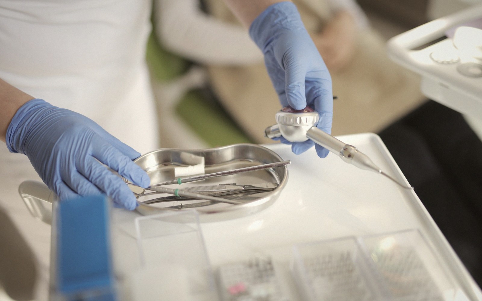
Once the animal is under and the airway is protected — a cuffed endotracheal tube keeps the mouth and throat separate, which matters when water and debris are in use — the vet or nurse begins with an ultrasonic scaler. This device uses high-frequency vibration and a water coolant to break down tartar from the enamel surface and from the sulcus, the groove between tooth and gum. Tartar is mineralised plaque; it harbours bacteria and, left in place, drives gingivitis and deeper periodontal disease. Scaling alone does not clean the root surfaces below the gum line, so hand instruments follow, working into the pockets where a probe has already measured disease depth. After scaling, the tooth surfaces are polished — a fine paste that smooths microscopic scratches left by instruments, because a rougher surface picks up new plaque faster.
No doses and no home remedies here: anything you give an animal should come from your own vet, for that animal.
Probing, x-ray and extraction
The probe is the key tool in any dental. It is a thin, blunt-tipped instrument that slips into the sulcus and measures pocket depth in millimetres. A shallow pocket — normal in dogs and cats — suggests a healthy attachment. A deep or irregular pocket, bleeding on probing, or a tooth that moves when pressed: all of these inform decisions. Every tooth is charted, which in a dog means working through forty-two teeth, and in a cat thirty. The result is a map of what the mouth looked like on that day, and it becomes part of the clinical record.
Dental radiography — intraoral x-ray — has transformed what vets can find. The visible crown of a tooth is roughly half of it; the root and the bone surrounding it are invisible to examination alone. X-ray reveals root fractures, bone loss, tooth resorption (common in cats), retained roots from previous extractions, and abscesses that have no external sign. Many practices now consider full-mouth radiography the standard for any dental, not a supplement reserved for obvious problems. It is the single tool most likely to change what happens in that session.
If x-ray or probing reveals disease that cannot be resolved by cleaning — a tooth with significant bone loss, a fractured root, or a lesion that has destroyed the attachment — extraction is the most common treatment. Veterinary extraction is more involved than the human equivalent for many teeth. Multi-rooted teeth are sectioned first: the crown is divided so that each root can be removed individually without levering against the bone. Gum flaps may be raised. The socket is curetted to clear any debris, then closed with absorbable sutures. For a tooth that has been doing real damage — a deep abscess, chronic pain from resorption, persistent infection — extraction resolves the problem rather than managing it.
Recovery from the anaesthetic happens in the practice, with monitoring until the animal is swallowing and responsive. Pain relief is given before the animal wakes, so that they are not in discomfort during the initial recovery period. Most animals eat within hours of going home, and owners are often surprised by how quickly behaviour improves — animals are skilled at concealing mouth pain, and a quiet, slightly withdrawn pet can turn noticeably brighter within a day or two of having a painful tooth removed.
What drives the cost
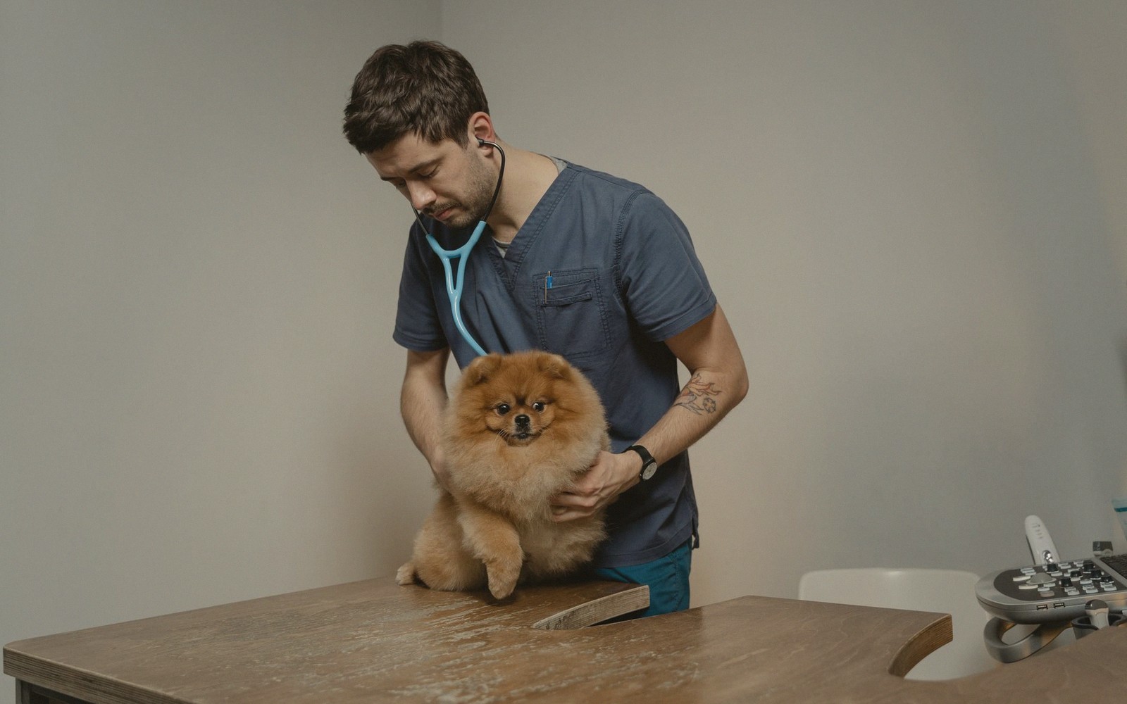
A dental is priced as a full anaesthetic procedure rather than a brief appointment, because it is one. The bill reflects pre-anaesthetic assessment, anaesthetic monitoring throughout, the time and instruments involved in cleaning, probing and charting every tooth, and radiography if taken. Extractions add to that, particularly complicated multi-rooted ones. It is common for an estimate to cover a range rather than a fixed figure, because the vet cannot know before the x-rays what they will find. Understanding that the range reflects genuine clinical uncertainty — not imprecision — makes it easier to work with.
Dental disease is extremely common in companion animals, and it progresses silently. Catching it at a routine check, before bone loss is significant, generally means less treatment in that session and a better outcome for the tooth. The mouth the vet examines at a booster appointment is one of the reasons the annual visit matters.