What Actually Happens in a Consultation
The history questions, the nose-to-tail examination, and what the vet's hands are actually reading.
The Questions Come First
Before a hand is laid on your animal, the vet will talk to you. This is not preamble — it is half the diagnosis. The history you provide shapes everything that follows, telling the vet where to look carefully and what findings to weight most heavily. A thorough history can be the difference between a sharp ten-minute examination and a longer, more exploratory one.
Expect to be asked when the problem started, whether it came on suddenly or crept in gradually, and whether anything has changed at home — a new food, a new pet, building work, a change in routine. You will be asked about appetite, thirst, urination and defecation, because those four together paint a remarkably complete picture of how the body's major systems are functioning. Vomiting and coughing questions are common too, and the vet will want to know the difference between something that happened once on Tuesday and something that happens every morning.
Vaccination status, parasite prevention, any current medications and previous health history will all be asked about. If your animal has seen another practice, or if you are coming in for a second opinion, bringing records saves time and avoids duplication. Jotting the key facts down the night before — dates, frequencies, anything you noticed — means nothing slips your mind when you are standing in the room and the clock is ticking. The details people most often forget under pressure are exactly the ones that matter most. Our piece on what to bring, and what to write down is worth reading before any appointment.
No doses and no home remedies here: anything you give an animal should come from your own vet, for that animal.
The Examination, from Nose to Tail
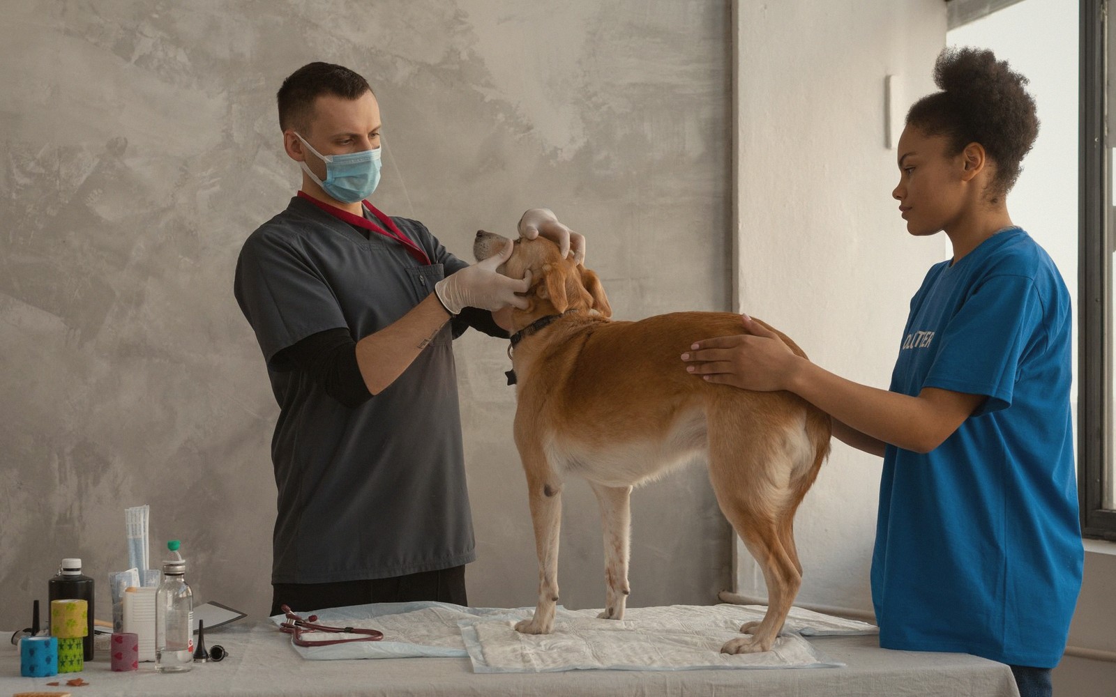
Once the history is taken, the physical examination begins. It follows a logic, not a ritual — the vet is working through body systems in a sequence that makes it hard to miss anything, even with a fractious patient or a consultation running short.
Starting at the head. The eyes are checked for discharge, redness, cloudiness or asymmetry. The ears are examined with an otoscope — a lighted scope that lets the vet see down the canal to the eardrum — and the vet is looking for inflammation, wax accumulation, foreign material and the smell that signals infection. The nose and mouth come next. Mucous membranes, usually assessed on the gums, give an immediate read on circulation and hydration: healthy gums are pink and moist, and when pressed briefly they refill with colour in under two seconds. Pale, white, blue or tacky gums are never normal and always warrant urgent attention. Teeth and gum margins are assessed — periodontal disease is widespread in companion animals and more consequential than most owners realise.
The lymph nodes. The vet's hands move to the neck, armpits, groin and behind the knees to feel the lymph nodes. These small glands are part of the immune system, and enlargement — either generalised or in one region — can be one of the earliest signs of infection, inflammation or something more serious. Most owners have never felt a lymph node on their animal; the vet does this every consultation.
Chest and cardiovascular system. A stethoscope goes to the chest. The vet listens to the heart on both sides, counting rate and rhythm and listening for murmurs — abnormal sounds caused by turbulent blood flow, which are graded on a scale from barely audible to heard without a stethoscope. Lung sounds are assessed at the same time: clear and even, or crackly, harsh and asymmetric. A healthy chest in a resting animal is quiet and regular. In older dogs and cats especially, this part of the examination sometimes finds things the owner had no idea were there.
The abdomen. Both hands are used here, and experienced vets can gather an enormous amount of information through the abdominal wall. The vet is feeling for the size, shape, position and texture of the major organs — liver, spleen, kidneys, bladder, and in female animals, the uterus. Intestinal loops should feel relaxed; tense, painful or distended loops are significant. The vet is also checking for fluid, masses and any response of discomfort when pressure is applied. In cats, kidney size and texture can be assessed quite directly through palpation, which is why kidney disease is often first suspected during a routine check rather than from owner-reported symptoms.
Skin and coat. The vet will part the fur and look at the skin itself — not just the topcoat. They are checking for fleas and flea dirt, scaling, redness, hair loss, thickening, lumps and the distribution of any lesion. Distribution matters enormously: a rash along the back is a different conversation from one confined to the paws or the groin. Any lumps found are assessed by feel — size, shape, whether they are attached to deeper tissue or move freely, soft or firm, painful or not. Fine-needle aspirates and biopsies are how lumps are characterised definitively, but the physical findings shape whether and how urgently that step is taken.
Limbs, joints and movement. Legs are flexed and extended, joints palpated for swelling, heat or crepitus, and muscles compared side to side for symmetry and bulk. In animals presenting with lameness, this part of the examination is extended and methodical. Muscle loss over one limb compared to the other is a sign of chronic underuse, pointing back in time to an injury or arthritic process the animal has been quietly compensating for.
Temperature, weight, body condition. Rectal temperature gives a baseline that anchors the whole picture — fever or hypothermia narrows the differential considerably. Weight is recorded and compared against previous visits, and most vets also assign a body condition score, a structured assessment of fat and muscle coverage that weight alone cannot capture. Gradual weight loss is one of the most commonly missed signs of underlying illness, because it is genuinely difficult to see at home when you live with an animal every day.
- 01 · The night beforeCarrier out, food withheld only if you were told to, and two lines written down about what you have noticed.
- 02 · Into the carrierThe top-loading trick, a blanket that smells of home, and why chasing a cat round a kitchen never ends well.
- 03 · The car journeyFootwell over back seat, cool air, and short practice runs that end somewhere pleasant instead.
- 04 · The waiting roomCarrier on a chair, not the floor. A towel over the door. Why the dog bench and the cat corner are apart.
- 05 · On the tableThe history questions, then a nose-to-tail examination: eyes, ears, mouth, chest, abdomen, joints, skin.
- 06 · Tests, and what they are forBloods, urine, x-ray, ultrasound. Which question each answers, and why some need sedation.
- 07 · Going home, and the billWritten instructions, a recheck date, and an invoice that reads differently once you know each line.
What the Vet Does with All of It
The examination takes as little as five minutes in a straightforward annual check, and considerably longer when the history is complex or the findings need unpicking. At the end of it, the vet will tell you what they found, what their working assessment is, and what they recommend next — whether that is reassurance, treatment, further tests or referral.
No single finding is usually conclusive on its own. What the vet is doing throughout is building a picture, triangulating between what you have told them and what their hands and instruments are reading. A murmur found in an otherwise well animal means something different from the same murmur in a dog who has been coughing at night. That synthesis — history, examination, context — is what a consultation actually is.
If anything found during the examination prompts further investigation, the vet will explain what they are looking for and why. Blood tests are one of the most common next steps, particularly where the examination suggests a systemic problem that cannot be resolved by physical findings alone.